
By Zachary Cronk, Physiotherapist
Understanding the vestibular system can be confusing! But it does not have to be. Just remember:
- Your vestibular system is a main contributor to your sense of balance and provides information about motion, head position, and spatial orientation.
- It is made up of several nerves, tracts and structures. It is typically thought to have a central (brain/nervous system) and a peripheral (inner ear) component that work hand in hand.
- Vestibular dysfunction can lead to things like vertigo or dizziness- which physiotherapy can help treat!
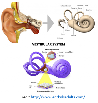 The main component that we will discuss today is your peripheral apparatus which is in the inner ear. It is comprised of a bony labyrinth that holds a membranous labyrinth with 3 semi-circular canals (SCC) and 2 Otolith organs.
The SCC consist of three tubes, that are filled with a fluid called endolymph. Towards the end of the tubes is a widening called the ampulla and within that is a sensory organ called the crista ampullaris, which is responsible for sending information about how we are moving in space. How Crista does this is by using information received from small hairs cells called stereocilia which extend out of the Crista into a membrane called the cupula- which acts as a barrier between the hair cells and the endolymph within the SCC. When the endolymph flows into the ampulla, it causes movement of the cupula, which then leads to the movement of the hair cells. This stimulates the hair cells which sends information to the brain about how the head moves.
The three SCC are positioned approximately at right angles to one another, they are each situated in a separate plane that will detect different movements: either a nodding movement, shaking head side to side or tilting left and right. These are designed to register rotational acceleration.
 The peripheral apparatus contains 2 other sensory organs called the otolith organs. These are critical for the detection of linear acceleration (forward and backward movements), gravitational forces, and tilting movements. The 2 organs are called the utricle and the saccule, they are responsible for detecting movement. The utricle is responsible for movement in the horizontal plane and the saccule is responsible for movement in the vertical plane.
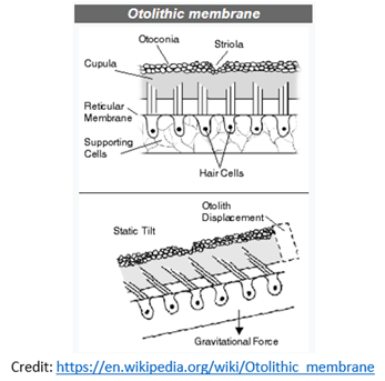
Within the otolith organs, there is a sensory organ called the macula. Similar to the ampulla of the SCC the macula also has hair cells. These hair cells extend into an otolithic membrane which is embedded with small crystals of calcium carbonate called otoconia. These crystals make the otolithic membrane heavier which means that when we move it causes the otoliths to sway and deflect the hair cells which send information about how we are moving in space. The structure and location of the Otolith organs make them sensitive to changes in linear acceleration and head tilts.
If you have any questions, please do not hesitate to book a session with one of our friendly physiotherapists. If you have any further questions, please contact us.
References
Lundberg, Y.W.; Zhao, X.; Yamoah, E.N. (2006). “Assembly of the otoconia complex to the macular sensory epithelium of the vestibule”. Brain Research. 1091 (1): 47–57.
Khan, S., & Chang, R. (2013). Anatomy of the vestibular system: a review. NeuroRehabilitation, 32(3), 437-443. S. M. Highstein, R. R. Fay, A. N. Popper, editors (2004). The vestibular system. Berlin: Springer.
Kingma, H., & Van de Berg, R. (2016). Anatomy, physiology, and physics of the peripheral vestibular system. In Handbook of clinical neurology (Vol. 137, pp. 1-16). Elsevier.